 |
||
Wasserman E.L.*, Kartashev N.K.**, Polonnikov R.I.**
Russia, Saint-Petersburg, Saint-Petersburg Institute for Informatics and Automation of Russian Academy of Sciences**, Saint-Petersburg V.M.Bekhterev Psychoneurological Research Institute of Public Health Ministry of Russia*, polon@mail.iias.spb.su
PROCESSING OF ELECTROENCEPHALOGRAMS OF CHILDREN WITH UNILATERAL BRAIN LESIONS BY MEANS OF ANALYSIS OF FRACTAL DYNAMICS.
Abstract. The results of express-processing of electroencephalograms (EEG) of ill and healthy children by means of evaluation of the shape characteristics of envelope of the current EEG power spectrums (the method of analysis of fractal dynamics) are described. On the basis of this processing automatic training procedures are built and the decision of assignment of EEG to the definite class is taken. The method can be used in telemedicine.
Вассерман Е.Л.*, Карташев Н.К.**, Полонников Р.И.**
Россия, Санкт-Петербург, Санкт-Петербургский институт информатики и автоматизации РАН**, Санкт-Петербургский научно-исследовательский психоневрологический институт им. В.М.Бехтерева МЗ РФ*, polon@mail.iias.spb.su
ОБРАБОТКА ЭЛЕКТРОЭНЦЕФАЛОГРАММ ДЕТЕЙ С ОДНОСТОРОННИМИ ПОРАЖЕНИЯМИ ГОЛОВНОГО МОЗГА МЕТОДОМ АНАЛИЗА ФРАКТАЛЬНОЙ ДИНАМИКИ.
Аннотация. Приводятся результаты экспресс-обработки электроэнцефалограмм (ЭЭГ) больных и здоровых детей методом оценки характеристик формы огибающей текущих спектров мощностей ЭЭГ (методом анализа фрактальной динамики). На основе этой обработки строятся автоматические обучающие процедуры и выносится решение об отнесении ЭЭГ к определенному классу. Метод может использоваться в телемедицине.
Subject. 51 persons with pre- or perinatal unilateral brain lesions resulted in hemiplegic cerebral palsy with an age range of 6-17 years (right hemiplegia (RH) ? 33 subjects, left hemiplegia (LH) ? 18 subjects) and 11 persons of the same age range with hemiplegia acquired in postnatal period of development (RH ? 4 subjects, LH ? 7 subjects) were examined during many-sided clinical, physiological and neuropsychological investigations by dr. E.L.Wasserman [1, 2, 3]. All subjects moved without assistance, served themselves in every day life and had normal or not severe disordered intelligence. The control group consisted of 47 practically healthy persons of the same age. Electroencephalographic (EEG) investigation was carried out in standard conditions with the use of common orientation and provoking tests, visual and mathematical analysis and numerical estimation of basic parameters. The scheme of placing of 12 “active” electrodes according to the system of R.Jung (1953) was used. The recording was carried out with the use of monopolar derivations and coupled ear electrodes as a “reference” one by means of 16-channel analogous EEG apparatus produced by firm Medicor (Hungary) with high frequency filter 30 Hz and time constant 0,3 sec. The signal was brought in IBM PC Pentium through 12-bit analog-digital transformer and then was recorded on the magnetic disk. The controlling of analog-digital transforming and of EEG monitoring during investigation as well as the recording on the magnetic disk were carried out with the help of original computer program elaborated in the laboratory of Child Neurophysiology of I.M.Sechenov Institute of Evolutionary Physiology and Biochemistry of Russian Academy of Sciences. Further processing of EEG was held in the conditions of postreal-time; the phone activity registered in passive waking state was analysed.
8 variants of classification of subjects under consideration were made according to clinical data and results of visual analysis of EEG:
Method. The processing of EEG was carried out by the method of analysis of fractal dynamics [4, 5] elaborated by prof. R.I.Polonnikov. This method assumes that parameters of system generating the fractal process (EEG) change in time and therefore it is possible to follow the dynamics of these changes by measuring the definite characteristics of fractal process at different time intervals. At that, the processing of EEG changes is more suitable to carry out not in time but in frequency field, because as it is known the power spectrum of fractal process is organized in definite order. This order is rather well described by two-parameter model of k*f-b form, where k ? coefficient characterizing power, f ? frequency and b ? informational parameter proportional to fractal dimension. The fractal dimension characterizes the speed of changing of information amount (according to R.V.L.Hartley). The frequency f is known, but k and b are subjected to estimation.
The time interval with duration in 1 second is chosen as the basic interval of time extension of EEG by which the power spectrum is calculated. The difference between the measured spectrum and the modelled one is the function of “leavings” which bring useful information about the shape of spectrum envelope in the frequency band of EEG rhythms as well as “trend” k*f-b . We investigated EEG fragmenton with duration in 30 sec. It means that the procedure of estimation of parameters repeated consecutively 30 times. Sufficiently complex statistical processing was carried out; as a result of it 9 informative parameters characterizing the changes of spectrums and allowing to make a judgement about dynamics of informative processes in organism were selected. The origin of these parameters is following: estimations b establish matrix with dimensions 30x12 (30 seconds and 12 derivations). This matrix allows to find integral average for b , normalized mean-square deviation and maximum singular number. Three similar parameters are obtained from matrix of k estimations and another three ? from matrix of leavings. On the basis of these 9 integral parameters the training procedure and the procedure of object recognition by the method of classical discriminant analysis with the use of Mahalanobis distance (method “Classify”) or by means of classical Bayes approach (method “Bayes”) were built. Selected 9 characteristics turned out to be universal and were used in any variants of partition in initial classes.
The processing program was written by N.K.Kartashev in MATLAB language (version 5.2). A number of M-functions for processing of EEG files and statistical analysis of results were developed.
function y=getind(name,beg,cnt) – calculates 9 classification characteristics according to EEG file; name – the name of processing file, beg – the number of the first count of digitised EEG, cnt – the amount of analysing counts. In our case we always analysed 6000 counts (30 sec.). Function returns the row vector of nine real numbers. Each element of it is a corresponding classification characteristic.
function crtab(num) – creates the training set; num – variant of partitioning in classes (1/2/.../8). Every training set is a set of matrices: the matrix of names of EEG files (name), the matrix of classes numbers (class) and the matrix of classification characteristics (tab). Function creates file containing these matrices.
function crsample(num) – creates the sample set. It is similar to crtab in usage and creates file containing matrices name, class, tab for sample set num.
For the processing of obtained tables two functions were used:
1. function predicted=classify(sample,training,class) – implements discriminant analysis (this function is included to Statistics toolbox of MATLAB);
2. function predicted=bayes(sample,training,class) – implements classifier according to Bayes method.
The both functions have same arguments: sample – matrix of coefficients of presenting objects; training – matrix of training set; class – column vector with the class number for every row of the training set. Functions return column vector with predicted class number for every row from sample.
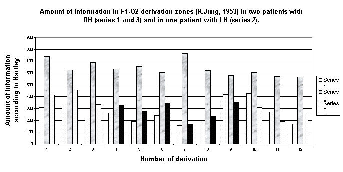
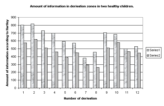
Results. The results of recognition are shown in table 1. Also the accurate correlation of several above-named characteristics with non-encephalographic parameters characterizing the abnormalities of every patient was discovered. At last, the dynamics of the first parameter (b ? estimation of EEG fractal dimension) is rather interesting. It is easy to pass from this estimation to the estimation of information amount according to R.V.L.Hartley. Fig. 1 shows the changing of the amount of information accumulated at 30 seconds in every derivation of EEG of ill child and fig. 2 ? of healthy child.
Table 1.
| Variant | Method “Classify”:% of
correct answers |
Method “Classify”: number of mistakes in I / II /III class |
Method of “Bayes”: % of correct answers |
Method of “Bayes”: number of mistakes in I / II /III class |
The whole of objects in given variant |
Number of objects in I / II / III class |
№1-training |
74,5 |
4/9 |
66,6 |
17/0 |
51 |
33/18 |
№1-control |
75 |
2/17 |
65,7 |
20/6 |
76 |
46/30 |
№2- training |
67,7 |
7/13 |
61,3 |
23/1 |
62 |
37/25 |
№2- control |
70,9 |
5/20 |
61,6 |
26/7 |
86 |
50/36 |
№3- training |
74,3 |
2/26 |
75,2 |
20/7 |
109 |
62/47 |
№3- control |
66,1 |
3/42 |
71,4 |
25/13 |
133 |
86/47 |
№4- training |
63,3 |
9/13/11 |
62,2 |
25/1/8 |
90 |
37/25/28 |
№4- control |
57,4 |
7/22/20 |
54,8 |
27/10/15 |
115 |
50/36/29 |
№5- training |
87 |
3/5 |
87 |
0/8 |
62 |
48/14 |
№5- control |
80,5 |
4/13 |
77 |
0/20 |
87 |
59/28 |
№6- training |
69,3 |
13/6 |
77,4 |
2/12 |
62 |
43/19 |
№6- control |
65,1 |
12/18 |
66,3 |
6/23 |
86 |
48/38 |
№7- training |
59,7 |
15/7/3 |
72,6 |
2/7/8 |
62 |
29/16/17 |
№7- control |
59,3 |
15/4/6 |
51,1 |
8/12/22 |
86 |
28/18/40 |
№8- training |
78,9 |
3/16 |
73,3 |
19/5 |
90 |
62/28 |
№8- control |
72,2 |
5/27 |
67,8 |
25/12 |
115 |
86/29 |
Comment: data obtained at following intervals with duration in 30 sec. repeated from one to three times in each case served as control selection.

Fig. 1.
Discussion. The use of any new method of obtaining and processing of information in medicine is always very interesting from the point of view of its possible diagnostic value. Automatic EEG classification in classes “norm-pathology” seems to be highly successful already in 70-80% of correct answers, because automatization of EEG analysis is regarded as one of the difficult tasks in clinical diagnostic. It is caused by high degree of complexity and nonstationarity of EEG signal as well as by empiricity of the classical clinical analysis of EEG. High percent (more than 80) of correct answers obtained during classification based on the result of visual estimation of fulfilment of functional activation tests was of especial interest since the EEG fragmentons corresponding to these tests were not included in the processing. So, it may be assumed that the used method allows to get the information concealed from the eyes of physician from the background brain activity; this information helps to forecast the character of work of central nervous system in the periods of information loading which is larger than at passive waking state with closed eyes.
Differences between brain regions in amount of accumulated information reflect probably by another language zonal differences conditioned by symmetrical gradient of main EEG rhythms in direction from front-to-back and successfully utilized as important diagnostic characteristic in clinical EEG. On the basis of the results of multivariate correlation analysis which discovered many links between 9 parameters under estimation and clinical and neuropsychological characteristics of brain work in our group of patients the usefulness and possible diagnostic value of the used method of EEG processing may be evaluated as optimistic, but it comes out of the framework of current report.

Fig. 2.
Comment to fig. 1, 2: 1-F2; 2-F1; 3-C2; 4-C1; 5-P2; 6-P1; 7-O2; 8-O1; 9-T2; 10-T1; 11-T4; 12-T3
Finally, applied method of EEG processing uses EEG fragmenton only in 30 seconds of duration and that’s why may be considered as express diagnostics and as we hope it will be useful in telemedicine.
References.
1. Wasserman E.L., Katisheva M.V. Multivariate clinical and neuropsychological study of higher mental functions in children with cerebral hemiplegia. // Bekhterev Review of psychiatry and medical psychology. — 1998. — № 2. — С. 45-52. [Вассерман Е.Л., Катышева М.В. Многомерное клинико-нейропсихологическое исследование высших психических функций у детей с церебральными гемипарезами. // Обозрение психиатрии и медицинской психологии им. В.М.Бехтерева] [Rus]
2. Katisheva M.V., Wasserman E.L. Neurological and neuropsychological investigation of children with unilateral brain lesions. // Brain and Development. — 1998. — V. 20., N. 6. — VIII Int. Child Neurology Congress, Ljubljana [Slovenia], September 13-17, 1998. Abstracts. — P. 447. — № 452.
3. Wasserman E.L. Clinical and morpho-functional correlations in hemiplegic cerebral palsy: Abstract of dissertation ... cand. med. science. ? Saint-Petersburg, 1999. [Вассерман Е.Л. Клинические и морфо-функциональные соотношения при гемипаретической форме детского церебрального паралича: Автореф. дис. ... канд. мед. наук][Rus]
4. Polonnikov R.I. Information measures at research of electrical activity of organism. // 55-the international seminar on Multimedia, Data Integration, Medical Databases. — Warsaw, 1999.
5. Polonnikov R.I. Information measures at research of biological processes. // Telemedicine ? formation and development: Materials of international scientific-practical seminar. ? Saint-Petersburg, Omega, 2000. — С.47-54. [Полонников Р.И. Информационные меры при исследовании биологических процессов. // Телемедицина – становление и развитие: Материалы международного научно-практического семинара] [Rus]
| Site of Information
Technologies Designed by inftech@webservis.ru. |
|